


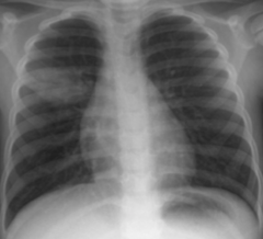
Pneumonia CXR

Lung Cancer CXR

RML.
Right heart border is being blurred/lost
Right heart border is being blurred/lost
Atelectasis CXR

This particular one is in the minor fissure
Loculated Pleural Fluid CXR

Pneumonia CT

Atelectasis CT

Lung Cancer CT

Pleural Fluid CT
The loss of a normal interface or border due to the pathological opacification of a region.
Silhouette Sign

Normal CXR Interfaces

-Accentuation of patent airways within an opacity
-Surrounding airspace if fluid filled
-More suggestive of pneumonia than atelectasis
-Surrounding airspace if fluid filled
-More suggestive of pneumonia than atelectasis
Air Bronchograms

-Peripheral opacity that rapidly evolves into a confluent homogenous consolidation
-Nonsegmental
-Effects entire lobe
-Commonly caused by streptococcus pneumoniae and klebsiella
-usually heals without sequela
-Nonsegmental
-Effects entire lobe
-Commonly caused by streptococcus pneumoniae and klebsiella
-usually heals without sequela
Lobar Pneumonia CXR
-Form of lobar pneumonia
-Klebsiella classically, S. Pneumo more common
-“Bulging fissure”
-Klebsiella classically, S. Pneumo more common
-“Bulging fissure”
“Round Pneumonia”

-Infection of the airway mucosa that extends into alveoli
-Patchy nodular opacities
-S. Aureus, or G- organisms
-Scarring after healing
-Patchy nodular opacities
-S. Aureus, or G- organisms
-Scarring after healing
Bronchopneumonia CXR
-Anaerobic bacteria
-Findings; bilateral medial lower low basal segment, right more common than left.
-Can become necrotic, capitate, and form an abscess.
-Any patient that cannot protect their airway is at risk.
-Findings; bilateral medial lower low basal segment, right more common than left.
-Can become necrotic, capitate, and form an abscess.
-Any patient that cannot protect their airway is at risk.
Aspiration Bronchopneumonia

-Viruses, M. pneumoniae, PCP
-Inflammation of interstitium
-Bilateral symmetric linear reticular opacities
-CT; Ground glass, whatever the &(%$ that is.
-Inflammation of interstitium
-Bilateral symmetric linear reticular opacities
-CT; Ground glass, whatever the &(%$ that is.
Interstitial Pneumonia
-Organizing pneumonia
-Cancer
-Timeline differentiates them.
-Cancer
-Timeline differentiates them.
Diseases that mimic pneumonia

-Disease with histo description of peripheral airspaces filling with mononuclear cells, foamy macrophages, and organizing fibrosis.
-Many known causes
-Findings; variable appearances with migratory multifocal peripheral opacities.
-Clinical; patient with protected nonproductive cough and low grade fever with restrictive pattern on PFT. Does not respond to antibiotics, does respond to steroids.
-Good prognosis.
-Many known causes
-Findings; variable appearances with migratory multifocal peripheral opacities.
-Clinical; patient with protected nonproductive cough and low grade fever with restrictive pattern on PFT. Does not respond to antibiotics, does respond to steroids.
-Good prognosis.
Organizing pneumonia

-Persistent opacity despite treatment
Cancer

-Get smaller post treatment
-Halo border
Acutely present or subside on serial imaging
-Halo border
Acutely present or subside on serial imaging
Infectious nodules

-Thick walled cavity
-Due to mixed anaerobic infection (S Aureus, pseudomonas)
-Often related to aspiration, poor dental hygiene, LOC, esophageal dysmotlity, neurological disease
-Due to mixed anaerobic infection (S Aureus, pseudomonas)
-Often related to aspiration, poor dental hygiene, LOC, esophageal dysmotlity, neurological disease
Lung Abscess

-Hematogenous spread of infection
-Multiple peripheral basilar nodules, which may cavitate.
-Some may show a feeding vessel, and an infarct
-Related to IVDU, and bacterial tricuspid valve endocarditis
-Staph Aureus and epidermis.
-Multiple peripheral basilar nodules, which may cavitate.
-Some may show a feeding vessel, and an infarct
-Related to IVDU, and bacterial tricuspid valve endocarditis
-Staph Aureus and epidermis.
Septic Emboli

-Purulent material in the pleural space
-Often related to evolution of a parapneumonic effusion, or an underlying lung infection that erupts into the pleural space (abscess or septic emboli).
-Often located
-Split Pleural sign
-Often related to evolution of a parapneumonic effusion, or an underlying lung infection that erupts into the pleural space (abscess or septic emboli).
-Often located
-Split Pleural sign
Empyema

-Granuloma; benign calcified nodules in the lung representing immune response to certain pathological insults.
-Caused by infectious and non-infectious causes
-Common infectious causes; Histo and TB.
-Often seen with calcified hilar/mediastinal lymph nodes and hepatic/splenic granulomata.
-Caused by infectious and non-infectious causes
-Common infectious causes; Histo and TB.
-Often seen with calcified hilar/mediastinal lymph nodes and hepatic/splenic granulomata.
Granulomatous disease in the lung

Progression of TB Infection
-Clinical infection following first exposure.
-Usually asymptomatic in children, only detected via PPD.
-Symptomatic in adults.
-FTT, night sweats, weight loss, hemoptysis.
-Often no imaging signs.
-Usually asymptomatic in children, only detected via PPD.
-Symptomatic in adults.
-FTT, night sweats, weight loss, hemoptysis.
-Often no imaging signs.
Primary TB

-Airspace consolidation, right more often than left.
-Mediastinal and ipsilateral hilar lymphadenopathy in children and immunocompromised. Atelectasis may occur from compression of central airways.
-Plural effusion, usually small, isolated, and unilateral.
-Findings clear slowly.
-Mediastinal and ipsilateral hilar lymphadenopathy in children and immunocompromised. Atelectasis may occur from compression of central airways.
-Plural effusion, usually small, isolated, and unilateral.
-Findings clear slowly.
Findings in primary TB
-Ghon complex; Visualization of sight of initial infection and enlarged ipsilateral lymph node.
-Ranke Complex; Calcified tuberculoma and ipsilateral hilar lymph node.
-Ranke Complex; Calcified tuberculoma and ipsilateral hilar lymph node.
Latent TB

-Consolidation process
-Extensive consolidation and cavitation can develop.
-Posterior upper lobe and superior segment of lower lobes is most common.
-Extensive consolidation and cavitation can develop.
-Posterior upper lobe and superior segment of lower lobes is most common.
Primary Progressive TB

-Reactivation TB
-Classically in the apical posterior upper lobes and superior segments of lower lobes.
-Rarely any pleural effusion or LAD.
-May be associated with Tree in Bud opacities, which indicates the spread of the disease via the small airways. (Image)
-Classically in the apical posterior upper lobes and superior segments of lower lobes.
-Rarely any pleural effusion or LAD.
-May be associated with Tree in Bud opacities, which indicates the spread of the disease via the small airways. (Image)
Post Primary TB

-Miliary TB
-indicates hematogenous spread
-indicates hematogenous spread
Disseminated Disease
-CD4>200; typical post-primary findings
-CD4<200; post primary resembles a primary infection; consolidation and LAD.
-CD4<200; post primary resembles a primary infection; consolidation and LAD.
Tuberculosis in the Immunocompromised
-Consolidation
-Endobronchial spread
-Miliary Patterns
-Centrilobular nodules (tree in bud)
-Primary, progressive primary, post-primary.
-Endobronchial spread
-Miliary Patterns
-Centrilobular nodules (tree in bud)
-Primary, progressive primary, post-primary.
Signs of Active TB
-Bronchiectasis
-Linear scarring
-Calcified nodules.
-Stable for 6mos.
-Linear scarring
-Calcified nodules.
-Stable for 6mos.
Signs of inactive TB
-M Avium Intracellulare Complex (MAC)
-From natural water, soil, and animals.
-Types; cavitary, bronchiectasis and nodules, centrilobular nodules.
-Symptom; chronic cough.
-From natural water, soil, and animals.
-Types; cavitary, bronchiectasis and nodules, centrilobular nodules.
-Symptom; chronic cough.
Non-Tubercular mycobacterium

-Resembles post primary TB
-Older men in 60s with COPD or mildly immunocompromised.
-Older men in 60s with COPD or mildly immunocompromised.
Cavitary MAC

-Bronchiectasis with waxing/waning nodules.
-Middle lobe and lingual predominant
-Women in their 60s.
-Lady Wndemere syndrome
-Middle lobe and lingual predominant
-Women in their 60s.
-Lady Wndemere syndrome
Bronchiectasis and nodules MAC

-Centrilobular ground glass nodules
-Owners of hot tubs
-“Hot tub lung”
-Owners of hot tubs
-“Hot tub lung”
MAC with hypersensitivity pneumonitis

-Bronchitis; cough and fever, +/- consolidation
-Bronchiectasis
-Bronchiectasis
Chronic Infection of the airways

-AR genetic disorder with decreased airway mucus clearance.
-Upper lobe in central cystic/varicoid bronchiectasis
-Pseudomonas, aspergillus, mycobacterial infection
-Upper lobe in central cystic/varicoid bronchiectasis
-Pseudomonas, aspergillus, mycobacterial infection
Cystic Fibrosis
-Invasive; neutropenic patients.
-Semi-invasive; mild immunocompromised patients. (Chronic necrotizing aspergillosis)
-Mycetoma; normal immunity, history of apical cavity.
-Findings; angio invasive (halos early, air crescent late), airway invasive (tree in bud and centrilobar nodules)
-Semi-invasive; mild immunocompromised patients. (Chronic necrotizing aspergillosis)
-Mycetoma; normal immunity, history of apical cavity.
-Findings; angio invasive (halos early, air crescent late), airway invasive (tree in bud and centrilobar nodules)
Aspergillosis
-Mild immunocompromised patients
-Chronic necrotizing aspergillosis
-Findings like TB; upper lobe consolidation and cavity.
-Chronic necrotizing aspergillosis
-Findings like TB; upper lobe consolidation and cavity.
Semi-invasive Aspergillosis

-Mycetoma.
-Normal immunity
-History of apical cavity (prior TB, bull, abscess)
Fungus ball fills a preexisting cavity.
-Normal immunity
-History of apical cavity (prior TB, bull, abscess)
Fungus ball fills a preexisting cavity.
Saprophytic Aspergillosis
